Tissue and tumor atlases revolutionize our understanding of disease progression and therapeutic response. They provide precise molecular data on cell types, states, and interactions in a preserved 3D environment to improve diagnosis and disease management.
Navigating Spatial Biology
The Harvard Tissue Atlas (HTA) gathers image and -omic datasets into high-resolution molecular maps. Our atlases provide precise molecular data on cell types, states, and interactions in a preserved 3D environment, shedding light on the complex interactions between cellular and acellular structures in normal and diseased tissue. These data provide a deeper understanding of how diseases start and progress to improve disease diagnosis and management.
The Harvard Tissue Atlas datasets enable a foundation for future advances in precision medicine, such as early cancer detection, AI/ML predictive models, and disease stratification for clinical trials.
Atlases


News


Publications & Software

Multiple human transgenes prolong survival of triple-carbohydrate knockout porcine kidney xenografts in nonhuman primates.
Explore Data

Highly multiplexed 3D profiling of cell states and immune niches in human tumours.
Explore Data
Publication
Multimodal spatial profiling reveals immune suppression and microenvironment remodeling in fallopian tube precursors to high-grade serous ovarian carcinoma.
Explore Data
Publication

AKT and EZH2 inhibitors kill TNBCs by hijacking mechanisms of involution
Publication
Featured Data Explorations
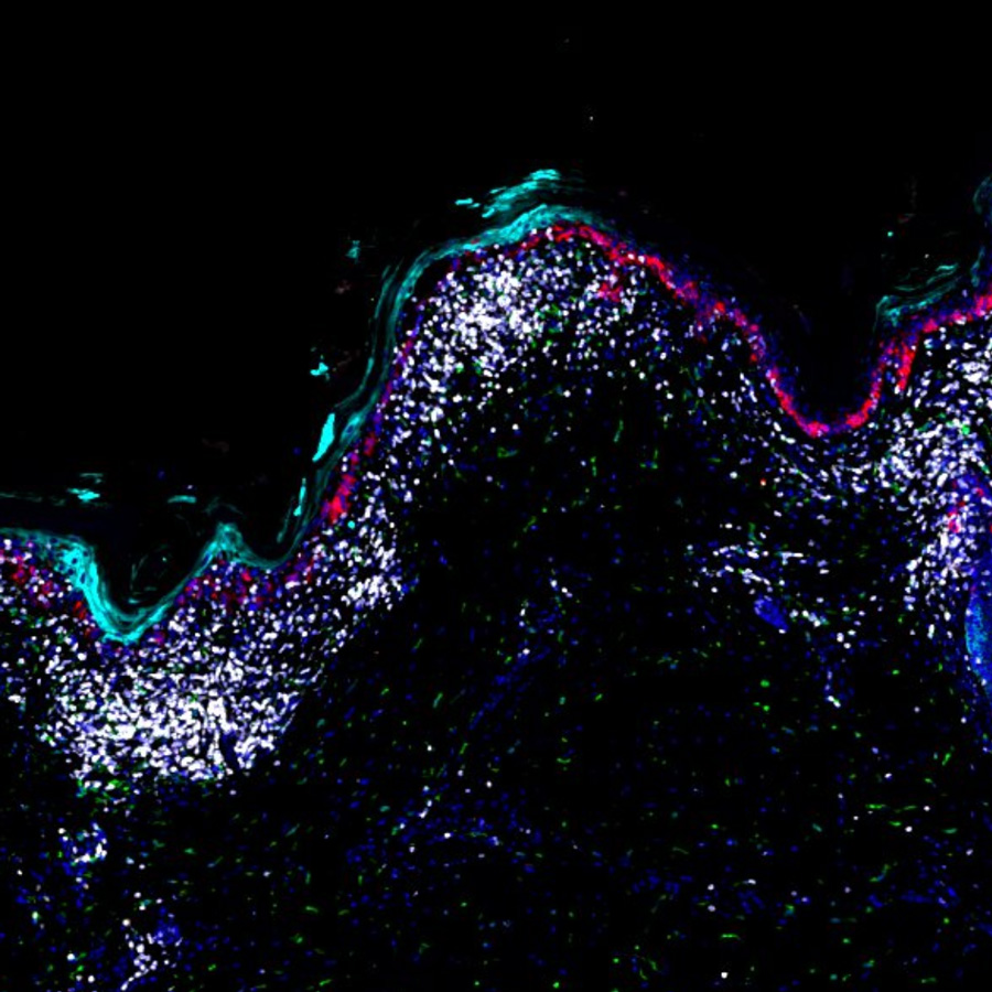
Skin immune-mesenchymal interplay within tertiary lymphoid structures promotes autoimmune pathogenesis in hidradenitis suppurativa - LSP25086

Melanoma in-situ with 3D imaging
Clear Cell Odontogenic Carcinoma (IF)
Ovarian STIC Spatial Transcriptomic Data from GeoMX and Multiplex Imaging
Multimodal spatial profiling of colorectal cancer using Orion
Graphical Abstract - Multiplexed 3D atlas of state transitions and immune interactions in colorectal cancer
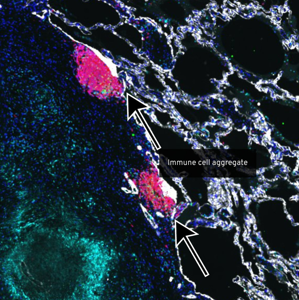
Exploring the Stages of Tuberculosis Granulomas
Patient 2 - Melanoma in situ, invasive melanoma, precursor field and inflammatory regression