Evolution of Delayed Resistance to Immunotherapy in a Melanoma Responder

Liu D, Lin JR, Robitschek EJ, Kasumova GG, Heyde A, Shi A, Kraya A, Zhang G, Moll T, Frederick DT, Chen YA, Wang S, Schapiro D, Ho LL, Bi K, Sahu A, Mei S, Miao B, Sharova T, Alvarez-Breckinridge C, Stocking J, Kim T, Fadden R, Lawrence D, Hoang MP, Cahill DP, Malehmir M, Nowak MA, Brastianos PK, Lian CG, Ruppin E, Izar B, Herlyn M, Van Allen EM, Nathanson K, Flaherty KT, Sullivan RJ, Kellis M, Sorger PK, Boland GM

Nature Medicine. 2021 Jun; 27: 985–992. PMID: 33941922.

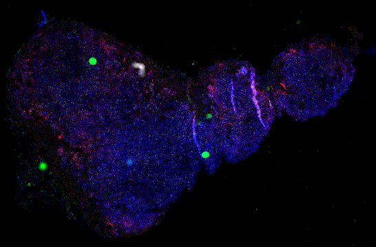
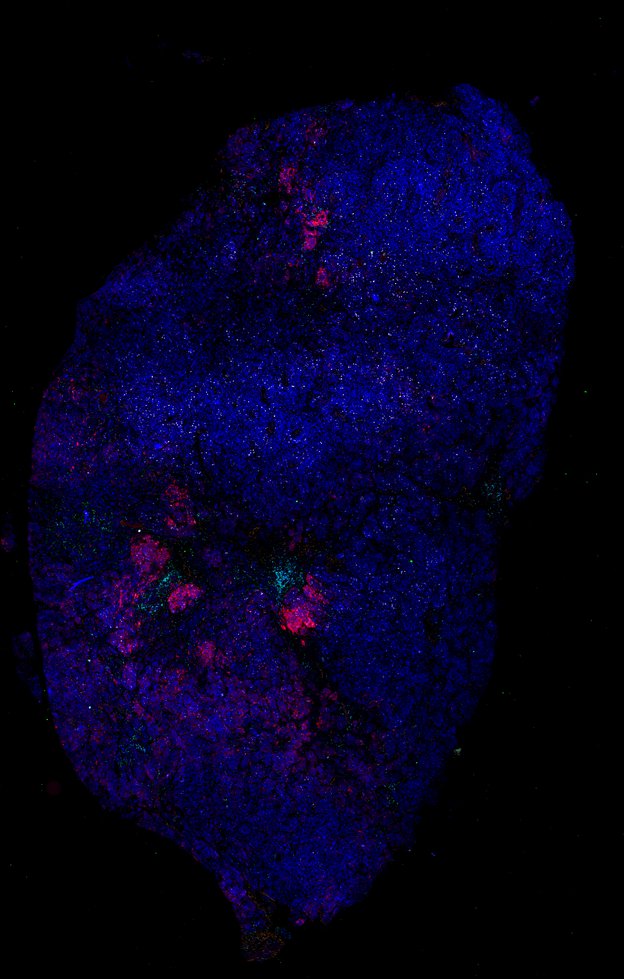
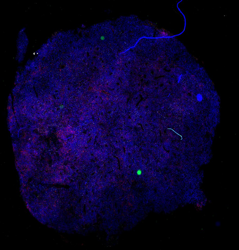
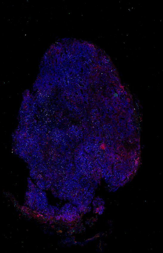
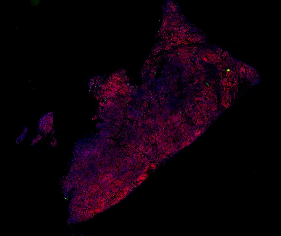
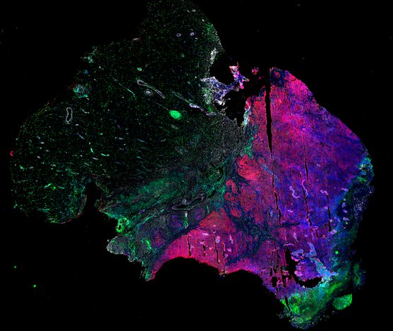
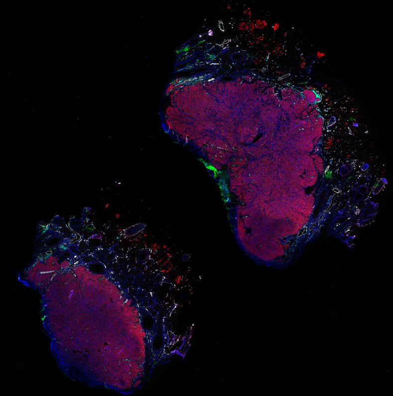
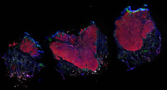
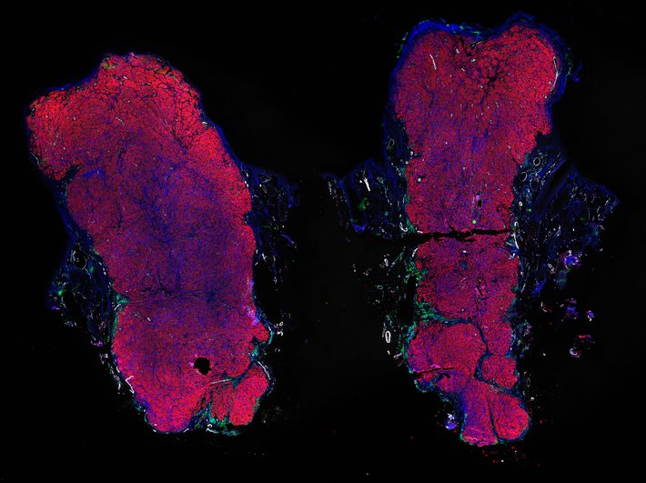
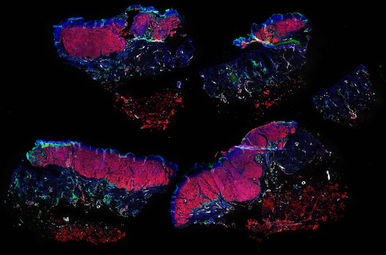
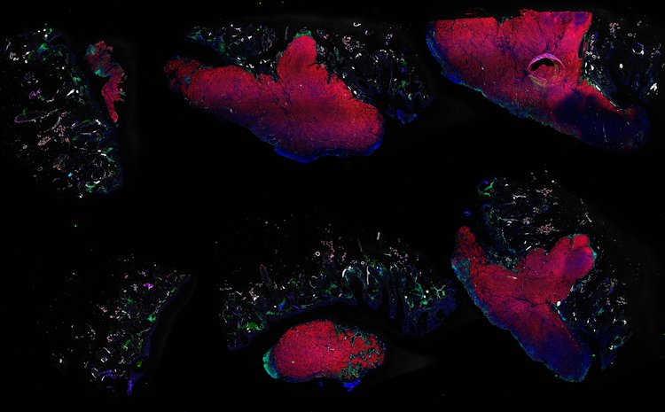
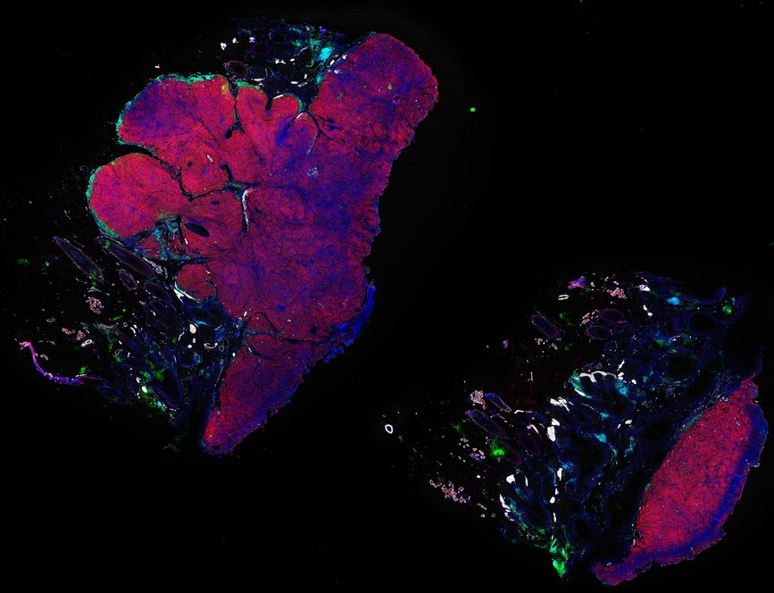
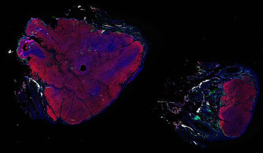
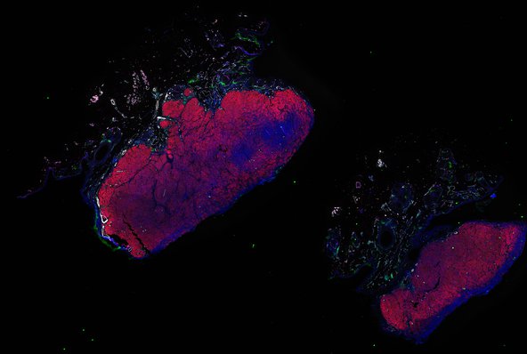
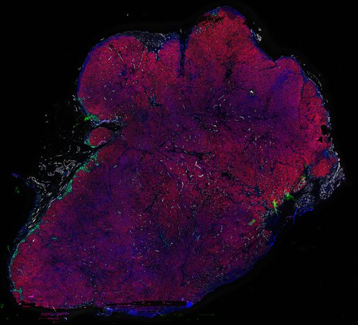
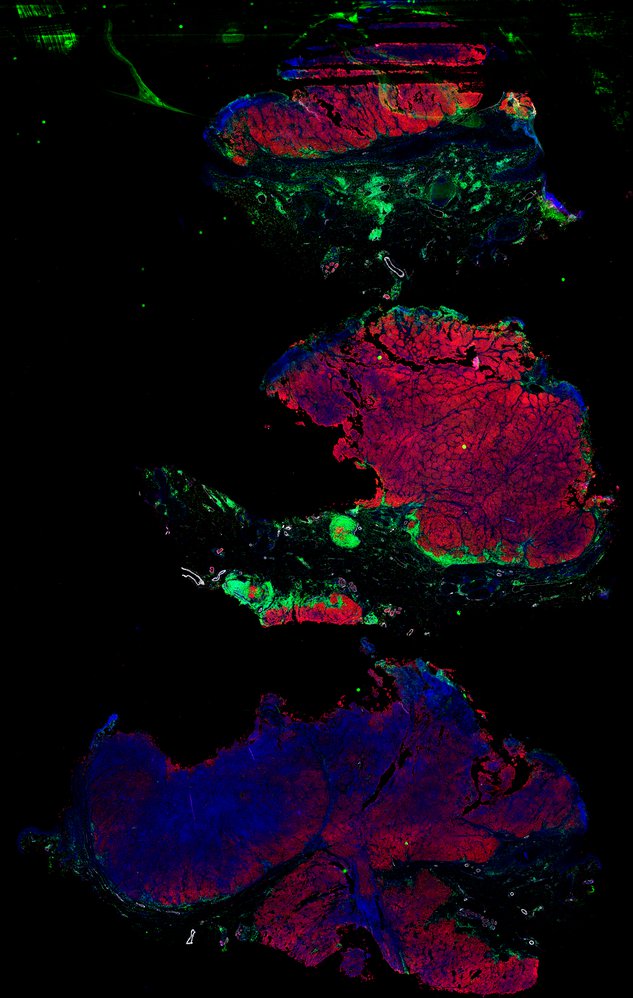
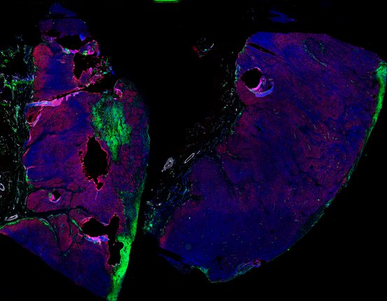
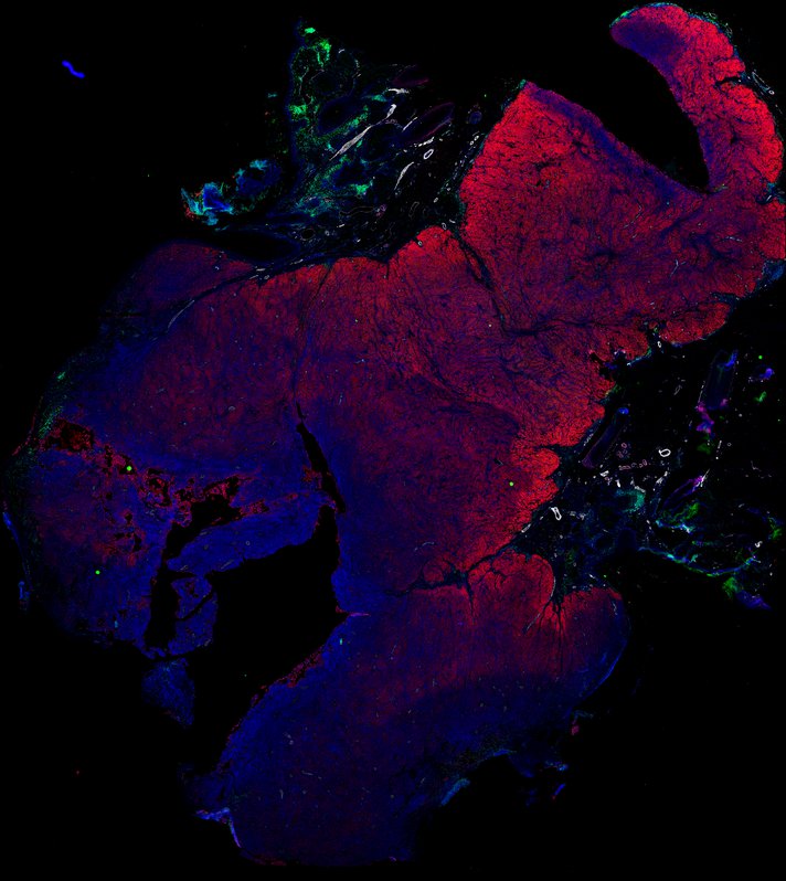
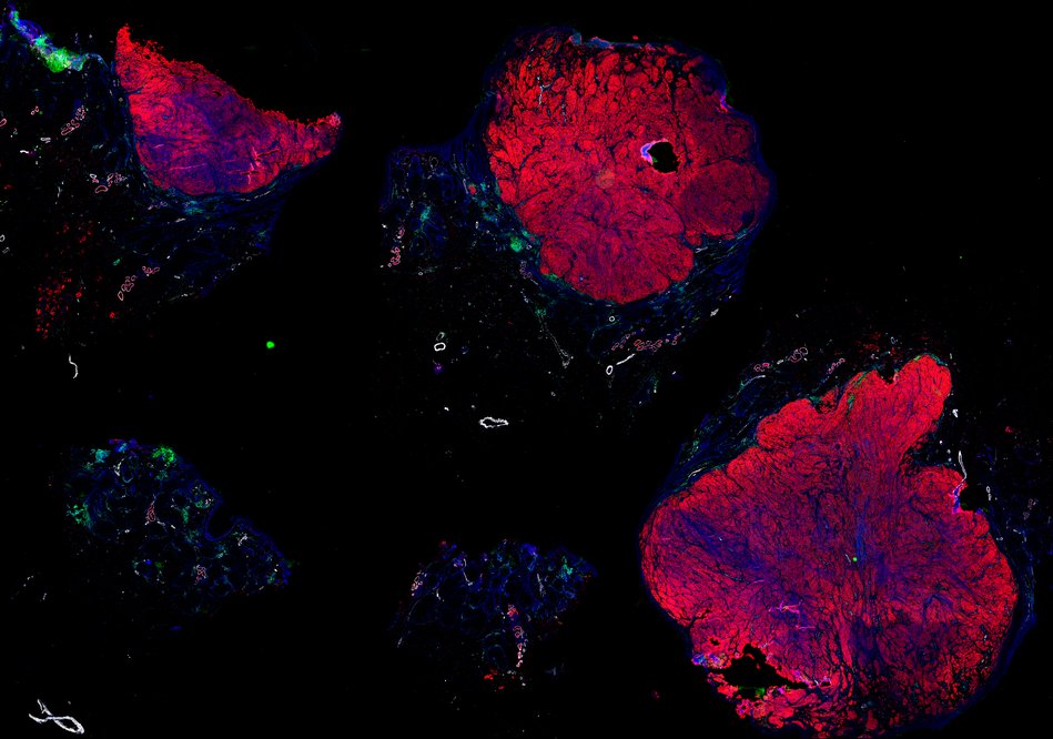
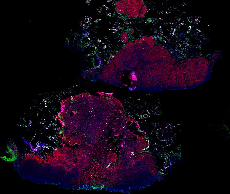
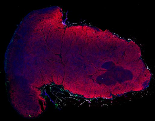
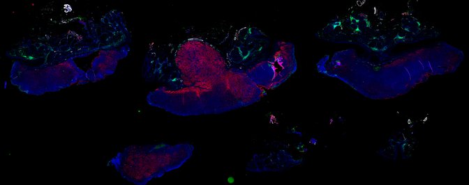
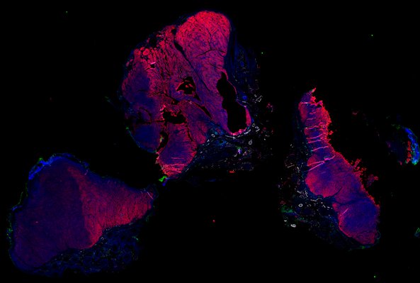
Despite initial responses, most melanoma patients develop resistance to immune checkpoint blockade (ICB) via poorly understood mechanisms. To understand the evolution of resistance, we studied 37 tumor samples collected over 9 years from a patient with metastatic melanoma and initial response to ICB followed by delayed recurrence and death. Phylogenetic analysis revealed co-evolution of 7 lineages with multiple convergent, but independent resistance-associated alterations (RAAs). All progression tumors emerged from a lineage characterized by loss of chromosome 15q including β2 microglobulin (B2M), with post-treatment resistant clones continuing to acquire additional genomic driver events including genome doubling, CDKN2A biallelic loss, and chromosome 11 loss. Deconvolution of bulk RNAseq and highly-multiplexed immunofluorescence (t-CyCIF) of histological specimens revealed differences in immune composition amongst the different lineages, despite an overall low immune composition. Imaging also revealed an NGFR-High tumor population enriched for PD-L1 expression in close proximity to immune cells with a vasculogenic mimicry phenotype. Rapid autopsy samples demonstrated 2 distinct NGFR spatial patterns with increased polarity and proximity to immune cells in subcutaneous tumors versus a diffuse spatial pattern in lung tumors, suggesting differences in the role and etiology of this neural crest-like program in different tumor microenvironments.
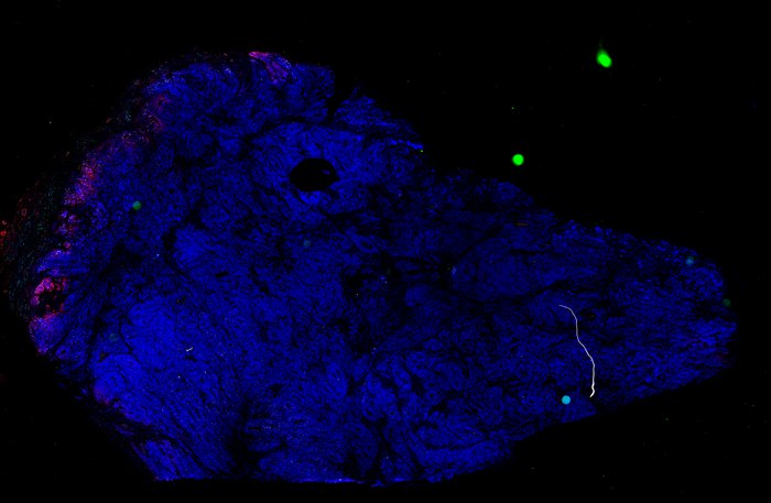
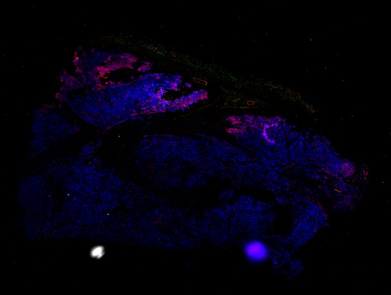
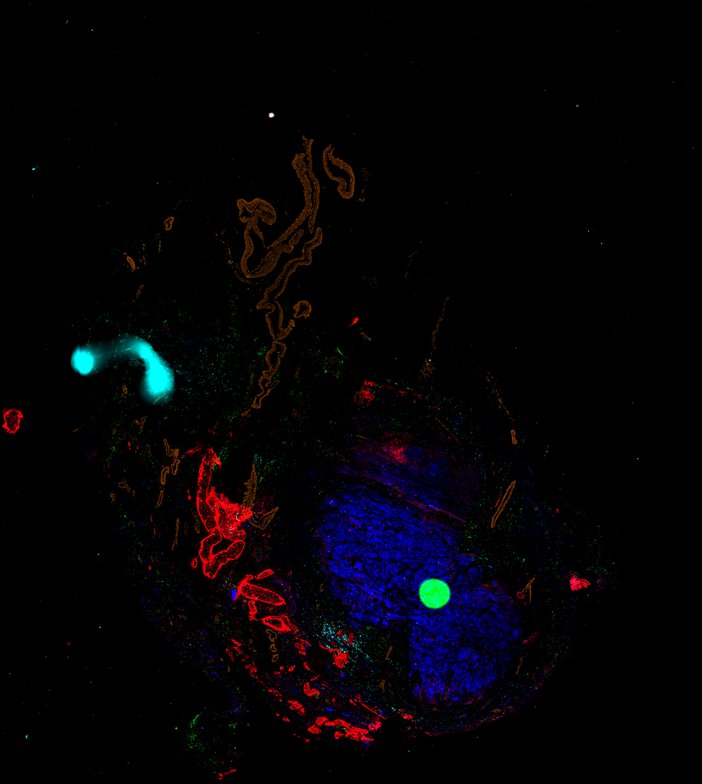
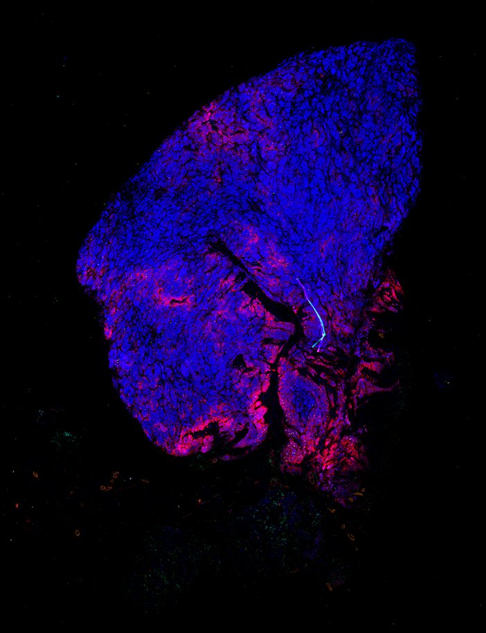
Publication Melanoma Atlas


timeline of patient disease progression, with treatments, and biopsies indicated Click to enlarge.


Curated Minerva Stories

Curated stories provide access to images that have undergone a quality control step to remove failed markers, ensure appropriate channel intensity settings, and provide metadata about the underlying sample and image. Click the Minerva story icon for an interactive view of the full-resolution images.

Data image
Longitudinal study of metastatic melanoma - Day 1849 - skin left neck (R3.1)
Data image
Longitudinal study of metastatic melanoma - Day 1862 - skin left neck (R3.2)
Data image
Longitudinal study of metastatic melanoma - Day 83 - skin left shoulder (T18)
Data image
Longitudinal study of metastatic melanoma - Day 109 - skin, left ring finger (T22)
Data image
Longitudinal study of metastatic melanoma - Day 1028 - Jejunal resection (R1)
Data image
Longitudinal study of metastatic melanoma - Day -22 - Skin, nasal labial fold (T4)
Data image
Longitudinal study of metastatic melanoma - Day 27 - Skin, upper lip (T6)
Data image
Longitudinal study of metastatic melanoma - Day 27 - Skin, chin (T5)
Data image
Longitudinal study of metastatic melanoma - Day 27 - Skin, left brow (T7)
Data image
Longitudinal study of metastatic melanoma - Day 27 - Skin, right ear (T8)
Data image
Longitudinal study of metastatic melanoma - Day 39 - Skin, right scalp (T9)
Data image
Longitudinal study of metastatic melanoma - Day 39 - Skin, left scalp (T11)
Data image
Longitudinal study of metastatic melanoma - Day 39 - Skin, posterior neck (T10)
Data image
Longitudinal study of metastatic melanoma - Day 39 - Skin, left arm (T12)
Data image
Longitudinal study of metastatic melanoma - Day 62 - Skin, right neck (T13)
Data image
Longitudinal study of metastatic melanoma - Day 62 - Skin, left wrist (T14)
Data image
Longitudinal study of metastatic melanoma - Day 76 - Skin, right neck (T16)
Data image
Longitudinal study of metastatic melanoma - Day 76 - Skin, right nasal labial fold (T17)
Data image
Longitudinal study of metastatic melanoma - Day 76 - Skin, left leg (T15)
Data image
Longitudinal study of metastatic melanoma - Day 92 - Skin, left groin (T19)
Data image
Longitudinal study of metastatic melanoma - Day 92 - Skin, left neck (T20)
Data image
Longitudinal study of metastatic melanoma - Day 92 - Skin, right upper lip (T21)

Automated Minerva Stories

Automated stories provide basic image viewing with automatic rendering settings, enabling rapid, lightweight sharing of highly multiplexed images, without download. Click the Minerva story icon for an interactive view of the full-resolution images.

Data image
Longitudinal study of metastatic melanoma - Autopsy - Lung, right lower lobe (AL1)
Data image
Longitudinal study of metastatic melanoma - Autopsy - Lung, right upper lobe (AL2)
Data image
Longitudinal study of metastatic melanoma - Autopsy - Lung, left upper lobe (AL3)
Data image
Longitudinal study of metastatic melanoma - Autopsy - Lung, left lower lobe (AL4)
Data image
Longitudinal study of metastatic melanoma - Autopsy - Subcutaneous lesion, left mastoid (AS1)
Data image
Longitudinal study of metastatic melanoma - Autopsy - Subcutaneous lesion, left inguinal region (AS2)
Data image
Longitudinal study of metastatic melanoma - Autopsy - Subcutaneous lesion, left scalp (AS3)
Data image
Longitudinal study of metastatic melanoma - Autopsy - Subcutaneous lesion, left temporal bone (AS4)
Data image
Longitudinal study of metastatic melanoma - Autopsy - Right adrenal (AV1)